
TMS Safety
Be aware of side effects
The risk of side effects can be minimised by screening patients for suitability for TMS using a TMS Safety Checklist. Potential side effects include syncope which is most likely due to anxiety rather than direct brain effects, TMS-associated seizure activity which is very rare for single-pulse TMS protocols, and transient headache or hearing changes (see the essential readings for more detail).
Not everyone can have TMS
The TMS Safety Checklist will cover absolute contraindications and safety considerations appropriate for the locality and ensure TMS can be used safely. The patient’s physician should be responsible for reviewing the checklist and deciding whether to approve the use of TMS. Very few patients (~2%) are unable to have TMS.
TMS operators should be properly trained by personnel with expertise in TMS
Training should include theory and practical sessions. Operators must be familiar with the means for summoning medical or emergency assistance in their facility. If seizure or syncope are suspected during testing, TMS should be terminated immediately and first aid delivered. It is advised that people who are pregnant should not operate TMS.
Module 1. TMS Overview
The mechanisms and physiology of TMS, and its potential side effects.
Overview
Transcranial magnetic stimulation (TMS) is a safe, painless, non-invasive way of stimulating neurons in the cortex of the brain
An insulated coil of wire is held gently against the scalp. A brief current is passed through this insulated coil, which generates a brief and rapidly changing magnetic field around the coil. The magnetic field passes painlessly through the scalp and skull. The magnetic field is shallow, and can induce current flow in the underlying tissues. This can depolarise neurons near the surface of the brain causing them to fire in unison.
The Motor System
The motor system consists of the central and peripheral nervous system and the muscles of the body, with the motor cortex the highest level of sensorimotor control.
The motor cortex is divided into several distinct regions including the primary motor cortex, premotor cortex, the supplementary motor area and the cingulate motor area.
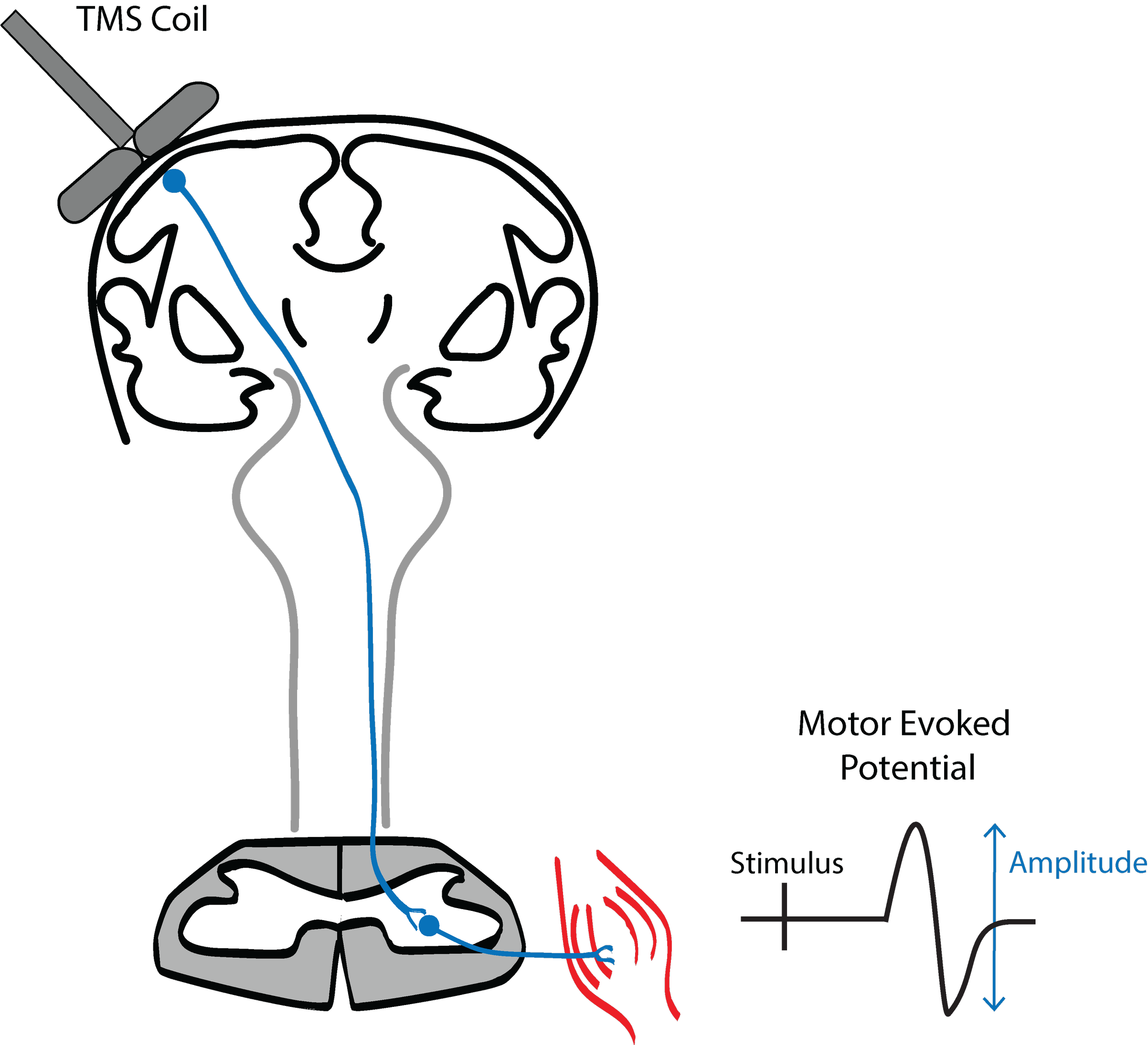
If the TMS coil is positioned over the primary motor cortex (located at the back of frontal lobe near the vertex) the magnetic stimulus can cause activation of pyramidal neurons that form the corticospinal tract. The pyramidal neurons of the corticospinal tract are excited by intracortical interneurons located in the more superficial layers of the cortex, layers 1 – 4. TMS excites the superficial intracortical interneurons which in turn cause synchronised firing of pyramidal neurons and descending volleys along the corticospinal tract.
Pyramidal neurons in the corticospinal tract cross the midline in the medulla before synapsing with alpha motor neurons in the spinal cord. When the synchronised descending output from the motor cortex activates alpha motor neurons, they fire action potentials that then travel along the peripheral nerves to muscles. Surface EMG can be used to detect the arrival of these action potentials in the muscles, observed as a motor evoked potential (MEP).
Obtaining MEP Status
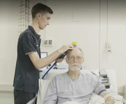
TMS can be performed at a patient’s bedside, in a procedure room, or similar location. Family members can be present during testing, if this is what the patient wants. For PREP2, testing takes about 20 minutes with the patient, with additional time before and after for setting up and putting away equipment, and documentation.
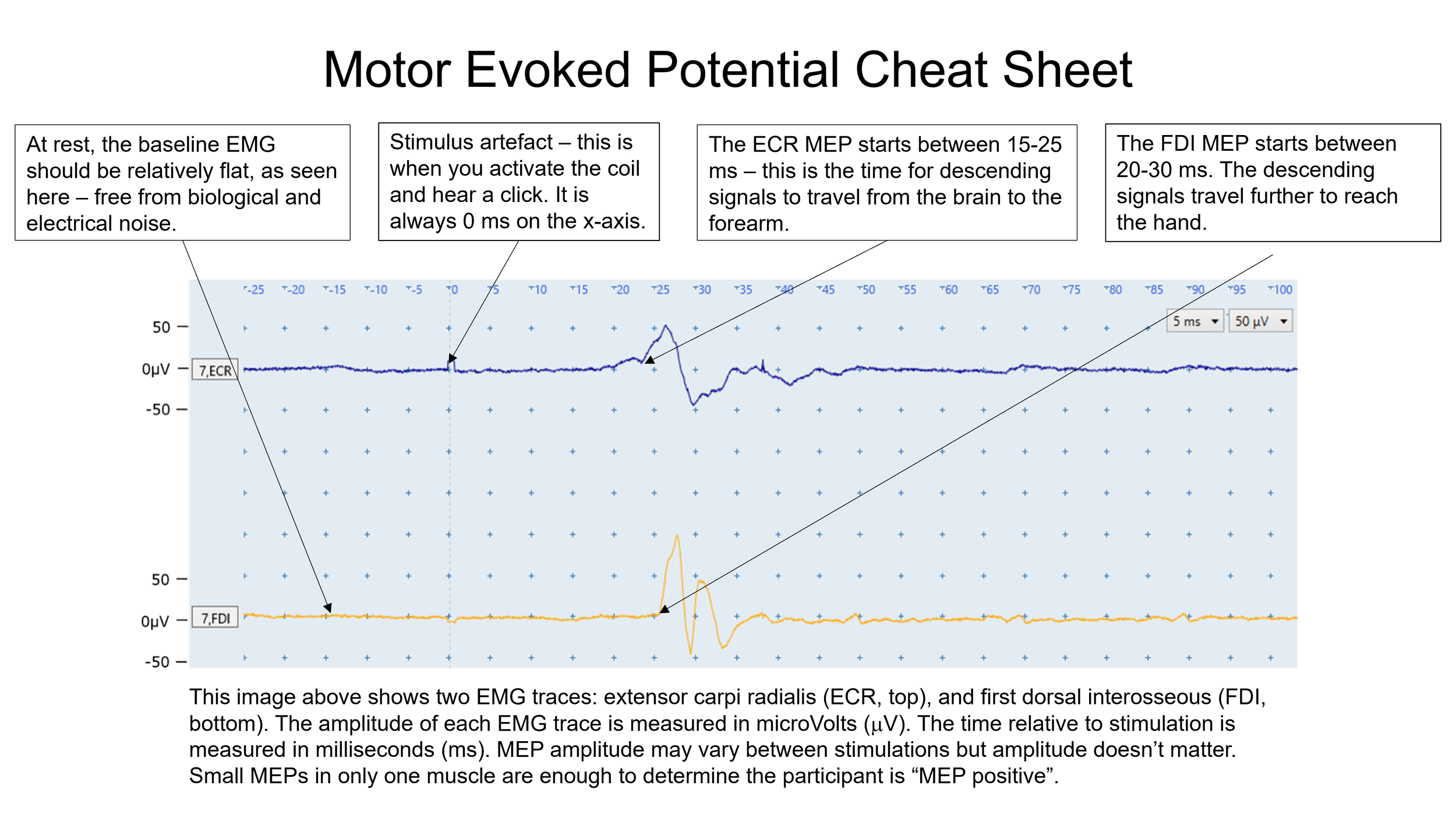
If TMS produces a visible MEP in the EMG trace, then the descending pathway from the primary motor cortex to the spinal cord is functional. The MEP status of the patient is MEP+.
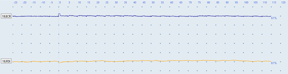
The picture below will help you orientate to key features of EMG traces that allow accurate determination MEP status. The picture shows an EMG recording from the extensor carpi radialis (ECR, blue trace) and first dorsal interosseous (FDI, yellow trace) muscles of the upper limb. If TMS produces a MEP then the descending pathway from the primary motor cortex to the spinal cord is functional. In this case the MEP status of the patient is MEP+. In the picture below you can see a MEP is present in both the ECR and FDI muscles and this patient would be classified as MEP+.
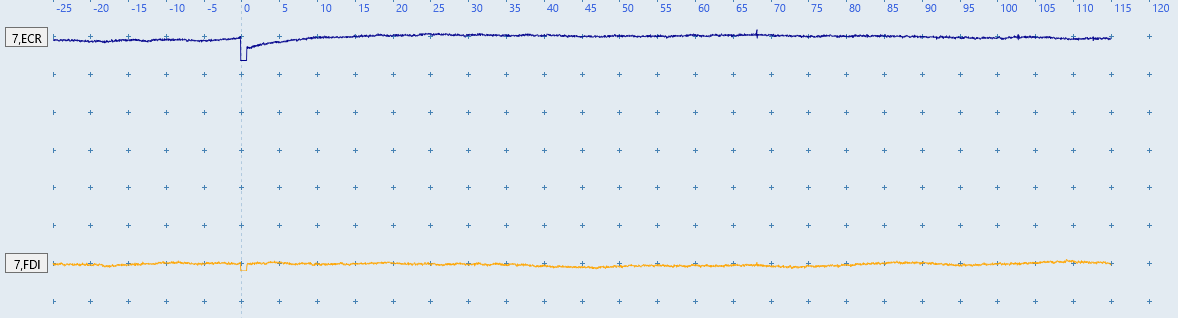
In the pictures below, EMG traces from two muscles show no MEPs. If this was representative of all TMS procedures and EMG traces for the patient, they would be classified as MEP-.
FAQs
Is it safe for everyone to have TMS?
1
People should be screened for contraindications to TMS to determine if it is safe for them before any testing takes place. You can screen patients with a TMS Safety Checklist, or an equivalent checklist already in local use. The patient’s physician should be responsible for reviewing the checklist and deciding whether to approve the use of TMS.
What does TMS feel like?
2
TMS is comfortable for most people. Before applying TMS, explain to the patient what they can expect. For example, when first describing TMS you might say:
"a plastic coil will be held against the side of your head to test the movement pathways from the brain to your weaker hand and arm"
"the device makes a click noise each time it is activated"
"you will feel a light tap on your head when the device is activated, but it shouldn't be painful"
"you may feel a brief twitch in your hand and arm muscles, but this won’t be painful. Not everyone feels the twitch, so it's okay if you don't feel anything"
Is single-pulse TMS like electroconvulsive therapy (ECT)?
3
Although both ECT and TMS involve stimulation of the brain their mechanisms, purpose, patient experience and safety profile are very different. ECT is a psychiatric treatment that involves directing an electric current into a patient’s brain via electrodes applied to the scalp to intentionally cause a series of generalized seizures. Patients are given muscle relaxants and general anaesthetic so they are not conscious during the treatment. Side effects such as memory loss are common.
By contrast TMS uses electromagnetic induction to non-invasively and painlessly activate cortical neurons. For TMS, patients are fully awake during testing and able to converse throughout the procedure. PREP2 uses single-pulse TMS which is an assessment tool, and not a treatment, and does not induce a series of seizures. Side effects are uncommon, and tend to be mild and transient.
If someone is due for TMS testing and their medical status changes what should I do?
4
Notify the person who is due to perform the TMS testing. They will need to review the situation and possibly revise the TMS Safety Checklist to reflect any changes. If the checklist answers change, then the physician who signed it off should be consulted to determine whether TMS testing is still appropriate.
Quiz
Complete the practice quiz.
Score at least 70% to receive the final quiz link by email.
Score at least 80% on the final quiz to receive a certificate of completion. Attempts are unlimited.